Course Directions
-
When you are finished reading the course & are ready to take the quiz, go to the bottom of this page & click the link underneath the Quizzes Section to begin the quiz.
-
After you complete the quiz with a passing score of 70% or higher, your certificate will be available to view & save for your records.
-
You have unlimited attempts to obtain a passing score on the quiz.
Helpful Tip
In case you need to double check an answer while taking the quiz, you can keep this reading material page OPEN by taking your mouse, and right clicking the “Quiz” link. Then, select the option to open to “Open Link in New Tab”.
This will open the quiz in a new tabbed window next to the reading material page.
Course Objectives
- Identify different mood disorders or types of depression
- Identify Hormones that play a role in depression.
- Identify what causes/contributes to depression
- such as chemical imbalances, and many other possible causes such as genetics, stressful life situations, medications, drug/alcohol abuse, and other medical issues.
- Identify different anatomical regions of the brain and how they affect mood.
- Define PET & explain how it works
- Define fMRI & explain how it works
- Evaluate research on the sizes of brain regions of people with depression and associated effects
- Identify the role of nerve communication and neurotransmitters in their connection with depression
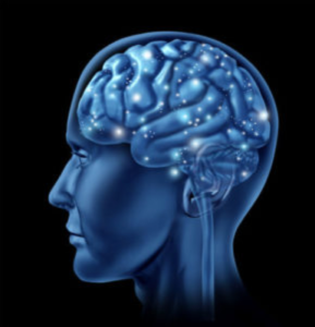
Depression can be a complex mental condition, and symptoms vary from person to person. To really understand this condition, it helps to be familiar with the part of the body where it almost all takes place, the brain.
The brain is a complex anatomical region with numerous functions and capabilities. This course aims to simplify different regions of the brain, associated hormones, neurotransmitters and the connection they have with depression so we can better understand this type of disorder from a more scientific perspective.
In the last section, we do a quick review on how and why acupuncture may be a useful therapy for abating depressive symptoms. By learning important anatomy and physiology of the brain, it can help provide us with a better understanding of individual personality, how depression arises, and how we can help.
Types of Depression
Before diving into the anatomy and science behind the brain, and how this connects with depression, we must first understand what depression is in the most basic sense. Depression is not a blanket term for all individuals because there are many varieties of depression. Hence, every patient is different and requires their own unique type of treatment. With a quick look in the DSM-VI, we can quickly see that there is more to this word “depression” than meets the eye.
Seasonal Affective Disorder (SAD)
This type of depression is known to affect people during the winter months, where the days are shorter and there is less sunlight. Luckily, the depressive feelings tend to lift in the spring and summer months. The root cause of mood changes may be from alterations in the body’s normal daily rhythms, or how chemical messengers like serotonin and melatonin function. One treatment for SAD is light therapy, which involves daily sessions sitting close to a very bright light source. Medication and/or psychotherapy may also be helpful, depending on the unique patient.
Persistent Depressive Disorder (dysthymia)
This type of depression is associated with low mood that has been persistent for at least two years but may not have reached the intensity of major depression. Some people with this disorder can function normally throughout the day, but feel down or unhappy much of the time.
Bipolar disorder
This type of mood disorder was once referred to as manic-depressive disease. It entails having episodes of depression. However, the individual usually goes through periods of strangely high energy or activity. The manic symptoms are the exact opposite of the depressive ones. The individual in mania may have very high self-esteem, grand ideas, decreased need for help, higher pursuits of pleasure.
There are four different types of bipolar disorder; Bipolar I, Bipolar II, Cyclothymia, Bipolar Disorder “other specified” and “unspecified.”
Mood Disorders that Lead to Depression
Several mood disorders that can lead to depression are bipolar disorder (manic-euphoric, hyperactive, inflated ego), persistent depressive disorder, cyclothymia, and seasonal affective disorder. People who tend to have addictive habits / behaviors and tendencies may be at higher risk for alcohol & drug abuse, including depression.
If a person has a history of a serious psychiatric disorder, they tend to have higher chances of having major depression. In addition, some people with major depression can show signs of anxiety and suffer panic attacks. Chronically anxious people may need to medicate themselves with either drugs or alcohol, and such habits can lead to depression.
The Brain & Personality

The brain governs all of our personality, and has many nerves that extend throughout our whole body. There are two types of neurons, sensory (afferent) neurons and motor (efferent) neurons. Sensory neurons take messages from the skin, tongue and other areas of the body and transmit them to the brain and spinal cord (Central Nervous System). Motor (efferent) neurons take information from the brain and transmit these important messages to the muscles or glands.
When it comes to depression, there is no one-size-fits all treatment to battle the condition because brain chemistry and functionality varies from person to person.
We also have diverse medical histories that need to be evaluated. For example, one person may have depression from a traumatic event during child hood, whereas another may have an unknown thyroid disorder that is causing his or her symptoms.
Each individual has a medical history specific to them, which may affect treatment outcomes. In addition, lifestyle choices may affect treatment. Due to the fact that everyone has different brain chemistry and genetics, different people may or may not respond well to certain treatments.
That being said, it is important to evaluate each patient as an entire person and to take a rigorous look at their medical history, current symptoms, and life-style.
Structures of the Brain
The Hypothalamus
The hypothalamus is defined as an area of gray matter, and has many functions throughout the body, and produces hormones that help regulate heart rate, sleep, mood, hunger, body temperature, thirst, sex drive, and more. This small region of the CNS is ultimately responsible for maintaining homeostatic functions in the body that are essential for survival. Among these important responsibilities of the hypothalamus are secreting specific hormones that do important things for our body.
Gonadotropin Releasing Hormone
This hormone helps to begin the onset of puberty, and travels from the brain to the pituitary gland, where it begins the production of two important hormones – luteinizing hormone and follicle stimulating hormone. Next, these hormones are released into circulation and act on the ovaries and testes to maintain reproductive functions. The importance of these hormones is significant because they control levels of hormones made by the ovaries and tests (i.e., progesterone, oestradiol, and testosterone).
Corticotrophin Releasing Hormone
This hormone is located throughout the brain and spinal cord, and can also be found in the GI tract, ovaries, testis, and pancreas.
Dopamine
When dopamine is released, it creates a feeling of happiness and well-being. This hormone also impacts motivation, emotions, learning, rewards, and sleeping, etc.
Oxytocin
This hormone is primarily associated with pregnancy and lactation, and has effects on the breast, uterus, and kidneys. It is also is known for its relaxing benefits to new mothers. Oxytocin is also released when there is physical connection between two people, in an interaction such as a hug or kiss. Also known as the “love hormone”, oxytocin is still being researched on its potential uses in helping depressed patients.
Vasopressin
Also referred to as ADH or Anti-diuretic hormone, vasopressin is associated with retaining water in the body, vasoconstriction, vascular smooth muscle hypertrophy, glycogenolysis, and platelet aggregation (Card, 2002). Similar to oxytocin, vasopressin acts on target cells in the breast, uterus, and kidney, but they do not stimulate the release of secondary hormones (Grimberg, 2017).
The ultimate goal of the hypothalamus is to keep the human biological system in homeostasis, by keeping fluid balances in check such as electrolytes, keeping body temperature within a normal range, monitoring blood pressure, and also working in our favor to help keep our bodies in a specific weight range.
Amygdala
The amygdala is located just posterior and superior to the hypothalamus, is a part of the limbic system of the brain, and has an important role in positive emotions (attraction, love) and negative emotions (anger, fear). The function of the amygdala is to link perceptions and thoughts about the world with their emotional meaning. In addition, this part of the brain governs the “fight or flight” response.
The amygdala contains many receptors for neurotransmitters, especially receptors for the stress hormone cortisol. Chronic stress may actually change the shape of certain brain regions because stress decreases the production of new neurons that grow in the brain.
According to research, the amygdala is highly active in individuals with depression, and it also plays a role in how we process negative and positive stimuli.
Hippocampus
The hippocampus is a part of the limbic system and has a role in one’s memory, retention of information, events, and emotions. The amygdala and hippocampus are connected in a few ways in their roles in the limbic system. The hippocampus enables us to gather sensory input from our outside world, link experience to emotion and to store all of it as coherent memories (Miller, Dudley, 2005).
In one study published by The Journal of Neuroscience, researchers found out that structure changes in the hippocampus occur in women who have recurrent major depression.
So, what does this mean?
The study ultimately found that the more episodes of depression the woman had, the smaller the hippocampal region in the brain. Some brain cells may be lost and others shrunken (Miller, Dudley, 2005). As mentioned prior, stress can play a role in this because of its ability to hinder the growth of new neurons.
Pre-Frontal Cortex (PFC)
This part of the brain is said to be the “executive part”, which helps us control our thoughts, emotions actions, working memory, planning, solving problems, and reasoning. In addition, this part of the brain has a lot of grey matter.
What is the significance of grey matter?
In the brain, there are two types of matter: Grey matter and white matter.
Grey matter contains mainly the neurons, and synapses, where all the messages in the brain are transmitted. Within the grey matter you will also find cell bodies, dendrites, and axon terminals. White matter on the other hand contains just the axons with the myelin sheath to speed up neuron transmissions.
The prefrontal cortex is crucial for cognition and the human’s ability to plan ahead, anticipate consequences, and emotional experiences like empathy.
In an interesting study, researchers found that individuals with major depressive disorder, who were able to “self-regulate” their negative emotions, had increased chances of recovering from depression.
The ability of the brain to self-regulate emotions takes place in the Pre-Frontal Cortex (PFC), and it is the PFC that then helps to regulate the amygdala and its emotional center (Heller, 2013).
Ultimately, it is the PFC that distinguishes your personality from everyone else.
Basal Ganglia
According to the research, the basal ganglia has a very important role in cognition reward and mood regulation. Abnormalities of the basal ganglia may play a role in the pathophysiology of mood disorders.
The basal ganglia are also interconnected with the thalamus, brain stem, and cerebral cortex. This structure is ultimately associated with emotion, learning, habits, cognition, voluntary motor movements, and eye movements. Dopamine also plays an important role in the basal ganglia.
The Neocortex / Frontal Lobes
The neocortex is the outer layer of the brain that is very unique to hormones. The frontal lobes (L & R) are important for “higher” cognitive functions like speech, complex reasoning, planning, and interpreting the world.
Electroencephalogram (EEG) studies, which measure the electrical activity of the brain, suggest that the left frontal lobes are more active when a person experiences pleasant emotions. Unpleasant emotions reflect stronger activity in the right frontal lobe.
The degree to which the 2 sides of the brain respond differently (brain asymmetry) may be an important individual difference associated with emotional sensitivity.
The importance of the frontal lobes also comes from the observed effects of brain injuries. Major injuries to this area can be very detrimental to the psyche and also, the overall functioning of the mind. Injuries to the pre-frontal cortex are even more damaging to developing children. Some examples of activities that could be problematic for this developing brain region are contact football or soccer (think head-butting soccer balls).
Neurons, Neurotransmitters, and Hormones
The nervous system is made up of cells, called neurons, which are connected with each other in complex ways. The brain is a thick bundle of neurons; other neurons form the brain stem and spinal cord (also known collectively as the Central Nervous System, or CNS), which connect the brain to muscles and sensory receptors, such as the ones on our skin, and all over the body.
Neurons are constantly communicating. The activity of one neuron may affect the activity of many other neurons, transmitting sensations from the far reaches of the body into the brain. The sensations are connected back to the brain, and may arouse feelings, memories, or action. Behavioral instructions are sent back out to the muscles, causing the body to move.
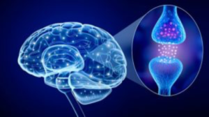
Communication between neurons is based on neurotransmitters. A bioelectrical impulse causes a release of neurotransmitters at the end of the neuron. These neurotransmitters travel across the synapse to the next neuron in line, where they cause a chemical reaction that results in that second neuron firing an impulse that causes the neurotransmitters at the other end to be released, and so on down the neural network.
Hormones, on the other hand, work slightly differently.
Hormones are released from central locations, such as the hypothalamus and the adrenal glands (on top of the kidneys), and spread throughout the body in the bloodstream. Once they reach the nerve cells that are sensitive to them, they will either stimulate or inhibit activity.
The difference between neurotransmitters and hormones can be confusing because they both affect the transmission of nerve impulses, and some chemicals qualify as belonging to both categories.
For example, norepinephrine functions as a neurotransmitter within the brain but is also a hormone that’s centrally released from the adrenal gland in response to stress.
Different neurotransmitters and hormones are associated with different neural subsystems and have different effects on behavior. For example, norepinephrine, dopamine, and serotonin work almost exclusively in the central nervous system (CNS)—the brain and the spinal cord. Epinephrine is found mostly in the peripheral nervous system (PNS)—the neuronal networks that extend all over the body.
Some neurotransmitters cause adjacent neurons to fire, while others inhibit the neuronal impulse. For example, endorphins work to inhibit, rather than promote, the neuronal transmission of pain.
Let’s look at the antidepressant Prozac. This medication increases the amount of serotonin in the body by inhibiting the activity of the chemical that causes the re-uptake (or removal) of serotonin. By inhibiting the chemical that essentially tells serotonin, “OK, you did your job, time to go”, the serotonin stays around, and continues to exert it’s pleasant, feel-good effects.
Epinephrine and norepinephrine are two particularly important neurotransmitters. Epinephrine and the neurons that respond to it are found throughout the body. Norepinephrine and its neurons work primarily within the brain itself.
Dopamine
Dopamine allows the brain to control body movements, reward response systems, and helps us approach attractive objects and people (also an important part of the chemical process that produces norepinephrine).
Dopamine plays a major role in depression. This hormone is said to be part of the basis of sociability and general activity level, and chronic low levels of dopamine (due to traumatic event or hormone imbalances) may cause an individual to be depressed.
An inherited inability of the brain to properly use dopamine can produce reward deficiency syndrome, characterized by outcomes such as alcoholism, drug abuse, and other complications. Additionally, a lack of dopamine in the brain may cause Parkinson’s disease.
Patients with Parkinson’s disease who took L-dopa (the precursor to dopamine) were able to have positive emotions again, motivation, sociability, and move around more. In addition, they also experienced interest and awareness of their environments. The dopamine systems in the brain may also play a role in bipolar disorder.
Dopamine may be relevant to the personality traits of extraversion and impulsivity. For example, an individual with less dopamine receptors is likely to be more tolerant of risk or dangerous activities. This is because when there are less dopamine receptors in the brain, it takes more of this chemical neurotransmitter to receive the same effect on the small number of receptors.
Neuropsychologist Jeffrey Gray hypothesized that dopamine and the structures in the hypothalamus are the basis of the tendency for desire to seek out reward by causing impulsive action. Dr. Gray refers to this as our “Go System.” Someone with a strong and active Go System will be more impulsive in his or her decisions.
This system interacts with other areas of the brain like the frontal lobes that assess and respond to risk and constitute a behavioral inhibition or “Stop” system.
Someone with a strong and active Stop system will tend to be inhibited and anxious. Because these are separate systems any combination of these two traits are possible.
Serotonin

Serotonin regulates the functioning of the endocrine and cardiovascular systems, and has an important job in the inhibition of behavioral impulses, regulating appetite, sleep, memory, mood, and body temperature. Low serotonin levels can have negative impacts on how a person feels and behaves. Some symptoms of decreased serotonin levels are having a hard time falling asleep, anxiety, memory and learning impairment, mood swings, and lastly, depression. People with insufficient serotonin have serotonin depletion. Other common symptoms include irrational anger, hypersensitivity to rejection, chronic pessimism, obsessive worry, and fear of risk taking. On the opposite end of the spectrum, excessive serotonin in the brain may also cause unwanted symptoms such as migraines and nausea.
Another word for serotonin is 5-hydroxytryptamine, a chemical substance that is derived from the amino acid tryptophan.
For individuals who have depression, researchers have postulated that there is an imbalance of serotonin levels that may influence mood. Low or imbalanced levels of serotonin could be from any one of the following reasons: a shortage of tryptophan, low brain production of serotonin, minimal receptor sites able to receive serotonin that’s being produced, or inability of serotonin to reach receptor sites (Bouchez, 2017).
According to the neuroscientist Dr. Alex Korb, serotonin activity can be boosted by remembering happy events, sunlight, exercise, and massage (Korb, 2011). Let’s take sunlight for example. In most cases, the UV rays from sunlight gets a bad reputation because too much exposure can cause skin cancer. However, UV light is vital for humans because when it is absorbed through our skin it produces vitamin D. Vitamin D in turn, helps with many positive functions in the body, including serotonin production. Additionally, compared to man-made light, natural sunlight occurs at the right time and helps us connect to our natural circadian rhythm.
Hormones
Hormones are biological chemicals that have effects on the body in a location different from where the chemical was produced. Some neurotransmitters such as norepinephrine can also be considered hormones because they affect nerve cells far from their origin.
Produced in a central location and broadcast throughout the body, stimulating the activity of neurons in many locations in the brain and body at the same time. Different hormones affect different nerve cells and influence different neural systems.
The hormones that govern behavior are released by the hypothalamus (part of the limbic system of the brain), the gonads (testes and ovaries), and the adrenal cortex (part of adrenal gland that sits on top of kidneys).
Testosterone

Testosterone is a known hormone that exists in both males and females but at different levels. Modern research shows us that women tend to, without their full awareness, be more attracted to men with higher levels of testosterone.
In one research study, women were asked to smell sweaty t-shirts worn by men who had just done a rigorous work out, and were asked to pick which t-shirt sample was “most appealing” or “tolerable” to them.
Researchers found that out of all the samples the women were given, that they chose the t-shirts of the men with the highest levels of testosterone. Males with more testosterone are higher in “stable extraversion”— sociability, self-acceptance, and dominance.
Higher levels of testosterone in women are associated with higher levels of self-reported sociability and impulsivity, lack of inhibition, and lack of conformity. Testosterone is not just a cause of behavior, it is an effect.
Here are a few interesting facts regarding hormones in females vs. males:
- Females produce 52% less serotonin than men according to one study, which may explain why men tend to adopt the “don’t worry, be happy” attitude more so than women. It may also may explain why women tend to experience more problems with worry, anxiety, and depression.
- Estrogen inhibits the neurotransmitter GABA, which helps relax and calm the brain.
- Testosterone boosts GABA which can lead males to feel less anxiety. When testosterone levels are at their peak in men or women (with polycystic ovarian syndrome) this can decrease levels of anxiety and exacerbate risk taking behavior.
- Anxiety disorders, including panic disorder, post-traumatic stress disorder (PTSD), social phobia, and generalized anxiety disorder, are more common in women.
Cortisol is released into the bloodstream by the adrenal cortex as a response to physical or psychological stress. Cortisol is part of the body’s preparation for action and is also an important part of several normal metabolic processes that can speed the heart rate, raise blood pressure, stimulate muscle strength, and metabolize fat.
The rise in cortisol seems to be an effect of stress and depression rather than a cause because injecting cortisol into people doesn’t produce these feelings. People with low levels of cortisol may be impulsive sensation seekers who are disinclined to follow the rules of society.
The levels of both hormones epinephrine and norepinephrine can rise dramatically and suddenly in response to stress or the “Fight-or-flight response”. In this case, the adrenaline starts to kick in, heart rate will go up, digestion processes are ceased, and muscles are tensed and ready for action. This is the sympathetic nervous system kicking in.
The brain becomes fully alert and ready to handle the task at hand. Almost all of the flight or fight studies have been conducted on males—animals and humans.
In contrast, a threat response may be handled differently for a woman carrying a child. Fighting or running may put her child at risk, so she therefore may come up with an alternative mechanism to protect herself and her child. For example, she may try to calm people down, or “tend and befriend” as some would say (Taylor, 2000).
Oxytocin is a hormone that has specific effects in females of promoting nurturing and sociable behavior along with relaxation and reduction of fearfulness. Strong feelings of trust will also activate the neurotransmitter oxytocin, in addition with several others such as dopamine and serotonin.
In contrast, when individuals feel threatened and distrustful, these happy hormones sharply decline and are replaced by norepinephrine, cortisol, and testosterone, preparing the person to either “fight or flight”.
This little part on “Fight or flight” is worth noting so we can see how our stress response should act normally. The stress-hormone system should kick in strongly during an emergency. For people who have depression, that system may remain switched on, especially for individuals who have had a very stressful childhood (Miller, Dudley, 2005).
Thyroid Disorders and Depression

There are many hormones that are produced by your thyroid, and imbalances of some of these hormones can lead to depression. The thyroid gland drives the production of serotonin, dopamine, adrenaline, and noradrenaline. Hypothyroidism, low thyroid hormone, can make you feel slow, tired, and sluggish throughout the day.
Not only that, but various bodily systems will be slow too, such as the digestive system, heart rate, and brain functioning. Research studies conclude that hypothyroidism ultimately lead to depression, decreased mental clarity, and anxiety.
When the thyroid is driving low production of the prior mentioned hormones, the adrenal gland hormones will try to compensate, especially with the hormone adrenaline, which can make the individual feel wired and hyper. On top of that, the levels of cortisol increase, increasing feelings of stress.
Many experts estimate that 1/3 of all depressions are directly related to thyroid imbalance. In addition, many people with thyroid imbalance may have impaired memory function.
Possible signs and symptoms for thyroid disorder are:
- Fatigue
- Weight gain
- Constipation
- Feeling cold
- High Blood Pressure
- High Cholesterol
- Dry eyes/skin
- Menstrual irregularities
- Infertility or recurrent miscarriages
- Thin/cracking/pealing nails
Research on the Brain & Depression
Dr. Helen S. Mayberg, Board Certified Neurologist and Professor of Psychiatry, Neurology, and Radiology at Emroy University School of Medicine in Atlanta, Georgia, has led an ample amount of research on depression and the brain, but a particular study conducted in 2005 produced some of the most significant findings about depression, psychiatry’s most common and elusive patient problem. Her research is highlighted below.
A decade prior to this particular research study, Dr. Mayberg had identified “area 25” in the brain, a key conduit of neural traffic between the thinking frontal cortex and the central limbic region that gives rise to emotion, which appeared earlier in our evolutionary development (Dobbs, 2006).
Many research studies she conducted found additional patterns. In the early 1990’s Dr. Mayberg and collaborators scanned 60 Parkinson’s patients, some depressed and others not, with positron-emission tomography (PET) (Dobbs, 2006). The goal was to find differences in activity in the frontal (the executive “thinking” region of the brain) and paralimbic regions (the emotion-governing region). They found that depressed patients showed far less activity in both brain regions (Dobbs, 2006).
Over the next couple years, Dr. Mayberg performed similar studies comparing depressed and non-depressed patients who experienced strokes or who had Huntington’s, epilepsy, or Alzheimer’s (Dobbs, 2006). She noticed the depressed patients in every study had the same reduced frontal and paralimbic activity (Dobbs, 2006).
In addition to these findings, area 25 was highly active. Now, depression is characterized by under-activity in the brain, so it was odd to find that one localized network was overactive (Dobbs, 2006).
Area 25 proved to have strong connections between the limbic system’s emotional and memory centers and the frontal cortex’s thinking centers. The researchers could not pin point exactly how area 25 regulated interactions between these two regions, but area 25 was glaringly alive in cases of severe depression (Dobbs, 2006).
Dr. Mayberg found that area 25 is highly active in individuals with depression, much more so compared to the other areas of the brain, which are vastly underactive. This specific area 25, since it is highly active, allows negative emotions to overwhelm thinking and mood.
Several other studies were conducted which ultimately showed that area 25 was highly active in all types of depression and was calmed by any successful therapy (Dobbs, 2006). Mayberg now had strong, replicated evidence that area 25 played a fundamental role in depression, and her insight had fit with what others had discovered about the factors of anxiety, stress, fear, and mood.
The researchers Joseph E. LeDoux, neuroscientist, and Bruce McEwan, a neuro-endocrinologist, had shown in their study that mood disorders often develop because of harsh or continuous stress. This could be from previous trauma or living in a difficult environment on a daily basis. These experiences can amp up fear and anxiety centers into the brain into “long term overdrive” (Dobbs, 2006). This survival system that has been permanently turned on in individuals with depression, is beneficial in extreme circumstances, but it becomes damaging when memories persist and thoughts trigger this system continuously (Dobbs, 2006). There is strong evidence for this dynamic, but the crucial switches in the circuit remained unclear (Dobbs, 2006).
Dr. Mayberg had thought that area 25 may be such a switch and tweaking it could get the circuit out of alarm mode and reduce the hyper activity to a normal area 25, which is as they had seen, is less active in individuals with no depressive symptoms.
So, she teamed up, at the time, with faculty members at the University of Toronto – a neurosurgeon Andres Lozano, and psychiatrist Sidney Kennedy.
The neurosurgeon Lozano had experience treating Parkinson’s patients by inserting next to the globus pallidus (a structure in the brain affecting individuals with Parkinson’s disease) a tiny, low voltage electrode. This technique is called deep brain stimulation, and it seemed to regulate the activity of the globus pallidus and thus restored movement in patients to near normal (Dobbs, 2006).
The researchers postulated… would inserting such electrodes alongside area 25 calm it down? (Dobbs, 2006) The team decided to try it.
“In 2003, the team implanted electrodes in area 25 in a dozen of severely depressed patients. Lozano drilled a pair of nickel size holes in the top of the skull, slid a pair of electrodes and slender leads to area 25, attached the leads to a small pacemaker sewn in under the collarbone and turned it on. The pacemaker sent a continuous four-volt current to area 25.” (Dobbs, 2006)
The results they found in their patients were amazing. According to the study, some patients felt profound relief as soon as the electrode was turned on, and 2/3 of the patient returned to essentially normal mood and function within months (Dodds, 2006).
Dr. Mayberg confirmed that area 25 causes depression when it’s hyperactive and calming it can bring relief (Dodds, 2006).
A Brief Overview of Anti-Depression Medications

Anti-depressants first started being prescribed for public use in the 1950s (Dudley, 2005). According to a psychiatric at the time, Andrew L. Morrison, luck played a small role in the discovery of this medication (Morrison, 1999). Originally, what became known as the anti-depressant drug Iproniazid was first being used to treat tuberculosis, until researchers found out it was alleviating some of the “downer” feelings of depression.
Iproniazid was the first that became known as a monoamine oxidase inhibitor (MAOI) (Dudley, 2005). Another class of anti-depressant known as tricyclics, was initially researched to be a cure for schizophrenia before approved as a treatment for depression under the brand name Tofranil (Dudley, 2005).
The effects of anti-depressant medications may not be felt until up to 8 weeks of treatment (Dudley, 2005). However, researchers and psychiatrists are really digging deep into the biology of depression and gaining new insights. In an interesting article by Michael C. Miller, assistant professor of psychiatry at Harvard Medical School and editor of the Harvard Mental Health Letter, new knowledge is discussed that researchers are amassing on depression.
The new model of depression doesn’t stem from a chemical imbalance of too little serotonin or too little norepinephrine, but instead from unhealthy or faulty nerve cell connections in the regions of the brain that create our emotions.
If that is true, and the evidence is robust enough, then the real goal of treatment isn’t to alter the chemistry of the brain by playing with levels of neurotransmitters, but instead to repair its circuitry,” (Miller, Dudley, 2005).
Here are some facts about the condition of depression and information on anti-depressant medications:
- Anti-depressants are only available by prescription from a doctor or psychiatrist.
- Major depression also carries the heaviest burden of disability among mental and behavioral disorders, according to the World Health Organization (WHO, 2010).
- Antidepressants were the third most common prescription drug taken by Americans of all ages in 2005-2008 and the most frequently were being used by people aging from 18 to 44 years old (National, 2010).
- Antidepressants adjust the way neurotransmitters work and interact within the brain, such as serotonin and noradrenaline.
- The effectiveness of such medications may be reduced if taken with alcohol or other drugs since both are metabolized in the liver (Dudley, 2005)
- According to the National Institute of Mental Health, major depression is one of the most common mental illness disorders in the United States (NIH, 2016).
- Overall, females are 2½ times as likely to take antidepressant medication as males. (NCHS, 2011)
Citations
American Thyroid Association, 2016.
National Center for Health Statistics. Health, United States, 2010: With special feature on death and dying. Table 95. Hyattsville, MD. 2011.
Lewis, T. (2013). Scent of a man.
Bloom, F.E. (2007). The Best of the Brain from Scientific American
Dobbs, D. (2006). Turning off depression. Scientific American Mind, Vol. 17, No. 4 August/September 2006
Mayberg, H.S., et al. 2005. Deep brain stimulation for treatment-resistant depression. Neuron 45 (5): 651-660.
Ashwell, K. (2012). The Brain Book.
Heller AS, Johnstone T, Peterson MJ, Kolden GG, Kalin NH, Davidson RJ. Increased Prefrontal Cortex Activity During Negative Emotion Regulation as a Predictor of Depression Symptom Severity Trajectory Over 6 Months. JAMA Psychiatry.2013;70(11):1181-1189. doi:10.1001/jamapsychiatry.2013.2430
Moritz-Saladino, A. (2015). 25 Facts you should know about grey matter.
Mental Health America
Healthline – Hypothalamus
Lafer B, Renshaw PF, Sachs GS. Major depression and the basal ganglia. (Dec 20, 1997). Phsychiatr Clin North Am.(4):885-96.
Taylor, S.E. (2000). Tend and Befriend.
McCarthy, L.A. Evolutionary and Biochemical Explanations for a Unique Female Stress Response: Tend-and-Befriend.
Dudley, W. (2005). A History of Drugs – Antidepressants.
Banov. M.D. (2010). Taking Antidepressants.
NIH. (2016). Major Depression Among Adults. 2015 SAMHSA NSDUH Mental Health Findings
WHO. (2010). World Health Organization.
NCHS Data Brief. (2011). Antidepressant use in persons aged 12 and over: United States, 2005-2008.
Medline Plus. (2018) Medical Encyclopedia. Hypothalamus
Card, J.P., Rinaman, L. (2002). Encyclopedia of the Human Brain. Hypothalamus. Science Direct.
Grimberg, A., Kutikov, J.K. (2017). Fetal and Neonatal Physiology (Fifth Edition).
Gonadotrophin-releasing hormone. (2017). You and your hormones.
Harvard Health Publishing. (2017). Six common depression types – ongoing mood, cognitive changes may require professional help.
Kevan Lee. Lifehacker (2014) How to Harness Your Brain’s Dopamine Supply and Increase Motivation
Course Directions
-
When you are finished reading the course & are ready to take the quiz, go to the bottom of this page & click the link underneath the Quizzes Section to begin the quiz.
-
After you complete the quiz with a passing score of 70% or higher, your certificate will be available to view & save for your records.
-
You have unlimited attempts to obtain a passing score on the quiz.
